


DENTAL PRACTICE WITH ELECTRONIC MICROSCOPE IN FLORENCE

ELECTRONIC MICROSCOPE
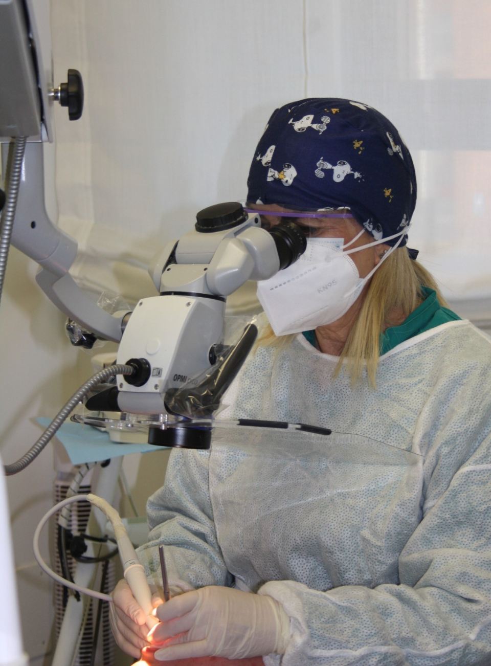
The electronic microscope has become a fundamental tool for modern dentistry. Being able to clearly enlarge and enhance the parts of the body that need to be observed makes it an essential tool when diagnosing problems and for the efficiency of the procedure, generating a change in practice which it would be difficult to think of turning back from. In addition to the magnification, what makes the real difference is the coaxial light beam that penetrates perfectly inside the area that needs to be looked at. There are countless areas of dentistry where using this kind of operating microscope has proved to be fundamental.
For example, at the
Baldi Dental Practice, the Opmi Pico Zeiss operating microscope is regularly used to carry out
oral hygiene, which gives a better but above all enlarged and detailed view of the surface of the tooth to be examined. This makes removing abnormal conditions and disorders more effective, such as bacterial plaque and tartar from the surface of the teeth.
This does not mean that if it is not used then the plaque removal has not been done successfully or as best as possible. However, the microscope is an effective way of helping to remove end the smallest, and most hidden or "invisible" agents.
It is also used to carry out endondontic treatments where the first treatment phase is actually the one that brings the most benefits, from opening the chamber to finding and looking into the canals. For example, in more complicated calcification of the chamber and calcified canal cases, we can only tell the calcific material from normal tissue (and turn neo-apposition dentin into primary dentin) and find the smallest traces of the position of the canal orifices by using a good magnification and lighting system
During surgery, we can plan to fit minimal invasive strips and use microsuture techniques. In restorative treatment, magnification can be used to ensure precise control of the areas to be restored.
In prostheses and prostheses on implants, the area of gum tissue that needs to be closed off can be checked and managed under the microscope.
Fill in our contact form to book an appointment


